
MCAT Organic Chemistry · Lesson 11
Spectroscopy
2 min read4 sectionsUpdated
4 sections
Spectroscopy Overview
Spectroscopy measures energy differences between quantized molecular states by analyzing absorption of electromagnetic radiation. Different types of motion (rotation, vibration, electron excitation, and nuclear spin transitions) produce characteristic spectra that reveal molecular structure.
- Measures energy differences between molecular states
- Types of molecular motion: rotation, vibration, electron absorption, nuclear spin
- Frequencies of EM radiation correspond to transitions between quantized energy levels
11.1 Infrared Spectroscopy
IR spectroscopy detects bond vibrations by measuring absorption of IR light. Characteristic absorption frequencies reveal functional groups and molecular fingerprint regions, particularly for polar bonds.
- Measures molecular vibrations: stretching, bending, or combinations
- Range: λ = 2500–25,000 nm → wavenumber = 4000–400 cm⁻¹
- Absorption occurs when bond dipole changes; nonpolar or symmetric bonds not detected
- Fingerprint region: unique to each molecule
- Characteristic absorptions:
- OH: broad ~3300 cm⁻¹ (alcohols), ~3000 cm⁻¹ (carboxylic acids)
- C=O: sharp ~1750 cm⁻¹
- N-H: sharp ~3300 cm⁻¹
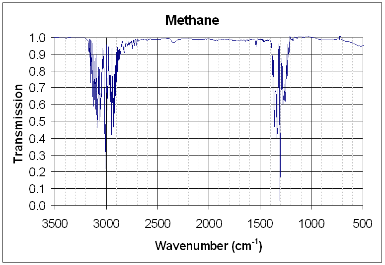
- IR spectra plotted as transmittance; max absorptions = valleys
- Useful for double/triple bonds and molecular fingerprinting
- Enantiomers have identical IR spectra
11.2 Ultraviolet Spectroscopy
UV spectroscopy measures electronic transitions of molecules with π or nonbonding electrons. Conjugated systems absorb longer wavelengths due to smaller HOMO-LUMO gaps, allowing identification of unsaturation patterns.
- Sample dissolved in inert, non-absorbing solvent
- Measures absorbance and λₘₐₓ
- Electronic transitions: π → π*, n → π*
- Smaller HOMO-LUMO gap → longer λ absorption
- Conjugation stabilizes excited states → shifts absorption
- Preferred method for conjugated alkenes
11.3 Nuclear Magnetic Resonance (NMR) Spectroscopy
NMR spectroscopy detects nuclear spin transitions in a magnetic field. Proton NMR provides information on hydrogen environments, chemical shifts, coupling patterns, and proton ratios, aiding structural elucidation.
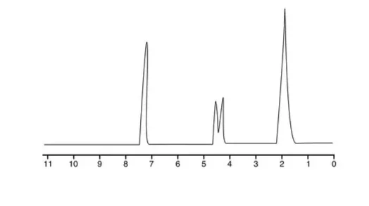
- Nuclei with magnetic moments align with (α, lower energy) or against (β, higher energy) B field
- MRI uses proton NMR for imaging
- Chemical shift (δ, ppm) plotted on x-axis, increasing left (downfield)
- Tetramethylsilane (TMS) = 0 ppm reference
- Proton NMR
- Typical range: 0–10 ppm
- Peak height ∝ number of protons
- Integration → ratio of protons (e.g., CH₃ vs HCl)
- Deshielding → downfield shift (electron-withdrawing neighbors)
- Shielding → upfield shift (electron-donating neighbors)
- Spin-spin coupling
- Occurs between protons 3 bonds apart
- N+1 rule: proton with n neighbors → split into n+1 peaks
- Coupling constant (J, Hz) measures splitting
- Multiplets = more than 4 shifts
- Chemical shift ranges (ppm)
- Aldehyde: 9–10
- COOH: 10.5–12
- Aromatic H: 6–8.5
- Higher electron density → more deshielded → more downfield
Spotted something wrong or unclear? Tell us — these notes are revised continuously.