
MCAT Biology · Lesson 7
Cardiovascular System
7 min read3 sectionsUpdated
3 sections
7.1 Anatomy of Cardiovascular System
The cardiovascular system includes a 4-chamber heart, vessels, and blood, with pulmonary and systemic circulation in series plus coordinated electrical conduction and valve-controlled flow.
- System has 4 chambered heart, blood vessels, and blood
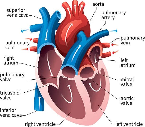
Heart
- 2 pumps, different circulations in series
- Right = pulmonary
- Left = systemic
- Venae cavae/pulmonary veins supply blood to atria
- Atria push blood into ventricles
Valves
- AV valves separate atria and ventricles
- Right = tricuspid valve
- Left = bicuspid (mitral) valve
- Semilunar valves separate ventricles and aorta
- Right = pulmonary valve
- Left = aortic valve
- 3 leaflets
Electrical conduction
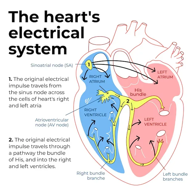
- Order of excitation:
- SA node → AV node → bundle of His → Purkinje fibers
- Starts at SA node → atria contract → ↑ atrial pressure (atrial kick)
- AV nodal delay allows ventricles to fill
- Bundle of His in interventricular septum → Purkinje fibers
- Cells connected by intercalated discs with gap junctions
- SA node intrinsic rate: 60–100 bpm
- Athletes: 40–50 bpm
- Autonomic control:
- Parasympathetic via vagus nerve
Contraction
- Systole = ventricular contraction
- Closure of AV valves
- Higher pressure
- Diastole = ventricles relaxed
- Semilunar valves closed
- Elasticity of arteries prevents pressure from falling
- Cardiac output:
- Usually
- Stroke volume (SV) = blood pumped per heartbeat
Vasculature
- Blood goes away from heart in arteries (aorta is largest)
- Pressure in aorta much higher than in superior vena cava
- Major arteries branch off aorta → further division
- Then branch into arterioles → capillaries
- Venous side:
- capillaries → venules → veins → vena cavae
- Blood vessels lined with endothelial cells:
- Maintain vessels
- Release chemicals that vasodilate/constrict
- Allow WBC passage
- Release chemicals for blood clots
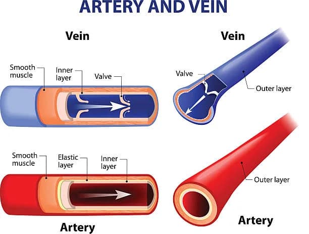
Arteries
- Most contain oxygenated blood
- Muscular arteries = arterioles
- Highly muscular + elastic → resistance to blood flow
- Heart must generate lots of pressure to overcome resistance
- Elastic recoil maintains pressure
Capillaries
- Single endothelial cell layer
- RBCs move single file → diffusion can occur
- Interface between circulation and tissues
- Allow endocrine signals to reach tissues too
- Bruise = leaking of blood from capillaries into ISF
Veins
- Thin-walled + inelastic
- Transport blood to heart
- Venules connect capillaries to larger veins
- Veins stretch to accommodate blood:
- ~ of blood is in veins
- Low smooth muscle → can stretch
- Total volume passing through either side of heart per unit time is same
Venous return + gravity
- Blood flow against gravity → more blood in vertical column
- High pressure in legs → veins have valves
- Varicose veins:
- Failure of venous valves → pooled blood
- Pregnancy ↑ risk:
- ↑ blood volume + compression of IVC by fetus
Skeletal muscle pump
- Skeletal muscle contraction squeezes veins → moves blood
- Why you should move → prevents blood clot formation
- Deep vein thrombosis (DVT) = clot in deep veins
- Can dislodge → pulmonary embolus
Circulation pathway
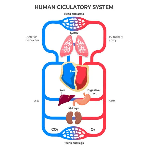
- Deoxy blood:
- vena cava → right atrium → valve → right ventricle
- pulmonary valve → pulmonary arteries → lungs
- pulmonary venules → pulmonary veins
- Oxy blood:
- left atrium → mitral valve → left ventricle
- aortic valve → aorta
- Blood usually passes through one capillary bed
- Portal system has two capillary beds
- Hepatic portal:
- hepatic portal vein → bed in liver
- Hypophyseal portal:
- bed in anterior pituitary → paracrine secretion → release hormones
- Renal portal:
- leaving glomerulus through efferent arteriole → goes to nephron
- Hepatic portal:
7.2 Blood
Blood is plasma plus formed elements (RBCs, WBCs, platelets), produced by hematopoiesis, and includes ABO/Rh antigen systems critical for transfusion and pregnancy.
Composition
- Blood is liquid and cells
Plasma
- Liquid portion
- Mix of nutrients, salts, respiratory gases, hormones, blood proteins
- Serum = plasma without clotting factors
- Preferred because lacks clotting factors + fibrinogen
Cellular portion
- Erythrocytes, leukocytes, platelets
- Formed from hematopoietic stem cells
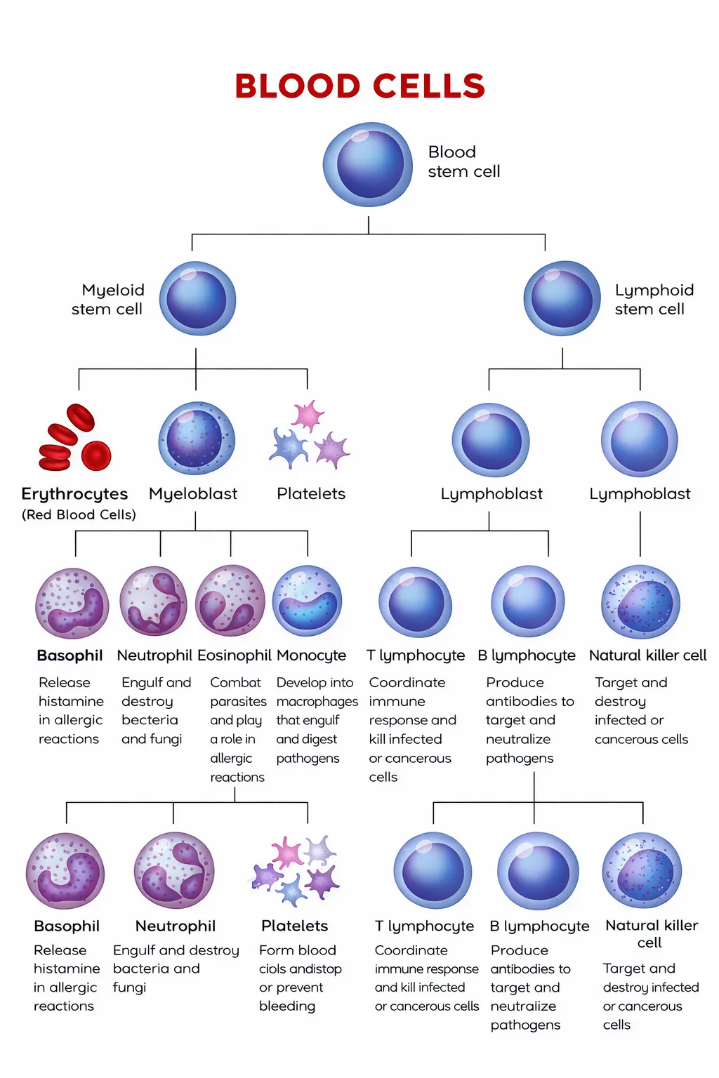
Erythrocytes (RBCs)
- Specialized for O₂ transport
- ~250 mil Hb per RBC, each Hb binds 4 O₂
- Each RBC carries ~1 billion O₂ molecules (as written)
- Biconcave:
- ↑ surface area
- Helps movement through capillaries
- Mature RBC has no nucleus → more room for Hb
- RBC doesn’t consume O₂ it carries:
- Uses glycolysis for ATP
- Lifespan ~120 days → then phagocytized (senescent RBCs)
- Hb = hemoglobin
- Hematocrit = % RBCs
- Hb ranges:
- Males: 13.5–17.5 g/dL
- Females: 12–15 g/dL
- Hematocrit ranges:
- Males: 41–53%
- Females: 36–46%
Leukocytes (WBCs)
- < of blood volume
- – per µL
- Defend against foreign invaders
- Granular leukocytes:
- Cytoplasmic granules visible on microscopy
- Toxic compounds released via exocytosis
- Inflammation, allergies, pus, bacteria destruction
- Agranulocytes:
- Lymphocytes + monocytes
Lymphocytes
- Specific immune response
- Primary responders
- Help body learn from experience
- Vaccines train immune cells via exposure to weakened pathogen
- B-cells mature in bone marrow, T-cells mature in thymus
Monocytes
- Phagocytize foreign matter
- Become tissue macrophages after leaving blood
- Tissue-specific names:
- Microglia (CNS)
- Langerhans cells (skin)
- Osteoclasts (bone)
Thrombocytes (platelets)
- Cell fragments/shards from megakaryocytes (bone marrow)
- Assist in blood clotting
- Present in high concentration
Hematopoiesis
- Blood cell + platelet production
- Triggered by hormones, growth factors, cytokines
- Erythropoietin:
- Secreted by kidney
- Stimulates RBC development
- Thrombopoietin:
- Secreted by liver + kidney
- Stimulates platelet development
Blood antigens
- RBC surface proteins = antigens
- Immune system reacts to antigens
- Major: ABO antigens + Rh factor
ABO antigens
- 3 alleles:
- A and B codominant
- Can have one, both, or none
- AB = A + B
- O is recessive:
- no A/B variant
- homozygous recessive genotype
- Must match blood type
- Severe hemolysis can occur if blood is foreign
- O⁻ = universal donor (no A/B/Rh antigens to trigger reaction)
- But O-type individuals make anti-A and anti-B antibodies
- AB = universal recipient (can take any type)
- Whole blood usually not transfused:
- packed RBCs (no plasma) are given
Antibodies
- Made in response to antigens
- No antibodies unless exposed
- Rh⁻ person does not naturally have Rh antibodies
- Gut E. coli proteins match A/B-like antigens:
- allows production of anti-A/anti-B antibodies prior to exposure
Rh factor
- Protein expressed on RBCs
- Several variants, but +/− refers to allele D presence
- Autosomal dominant inheritance
- Maternal-fetal medicine:
- Rh⁻ mom + Rh⁺ fetus → sensitization → antibodies made
- Subsequent Rh⁺ fetus → antibodies attack fetal RBCs
- Erythroblastosis fetalis (can be fatal)
- ABO mismatch less concern (antigens can’t cross placenta)
- Rh IgG antibodies can cross placenta
7.3 Physiology of Cardio System
Cardiovascular physiology depends on maintaining blood pressure and flow, exchanging gases/solutes at capillaries, regulating fluid via Starling forces, and preventing bleeding through coagulation.
Blood pressure
- Must be kept sufficiently high
- Force per unit area on vessel walls
- Measured with sphygmomanometers
- Normal: 90/60 and 120/80 (as written)
- Relationship:
- Arterioles and capillaries act like resistance
- Arterioles can constrict → ↓ flow, ↑ resistance
- Systemic capillary beds in parallel (except portals)
Regulation
- Baroreceptors regulate BP
- Too low BP:
- ADH/vasopressin
- Low perfusion to JG cells → renin → aldosterone → Na⁺ reabsorption → more water
- Too high BP:
- ANP released
Gas and solute exchange
Oxygen
- Carried by Hb:
- protein with 4 subunits + prosthetic heme
- Blood O₂ level measured by :
- – mmHg
- O₂ saturation:
- Cooperative binding:
- O₂ binding increases affinity (easier for more O₂ to bind)
- O₂ leaving decreases affinity (easier for more to leave)
- Affinity decreases as pH decreases
- During exercise:
- mmHg (as written)
- normally mmHg (as written)
CO₂
- Mostly as bicarbonate
- Carbonic anhydrase catalyzes conversion
- ↑ → ↑ protons → ↓ pH
- Protons reduce Hb affinity for O₂ → right shift (Bohr effect)
- Trigger usually tied to O₂ demand
- Bicarbonate system also buffers
Left shift from
- Decreased
- Decreased
- Increased pH
- Decreased temp
- Decreased 2,3-BPG
- Fetal HbF has higher affinity than HbA
- Fetal cells pull O₂ off maternal cells
Kidney + acidosis
- Kidneys account for some acidosis handling
- Renal tubular acidosis type I:
- can’t excrete acid effectively
- proton buildup, buffer shifts left
Nutrients, waste, and hormones
- Carbs + AAs absorbed into small intestine capillaries → hepatic portal system
- Fats absorbed in lacteals → enter via thoracic duct
- Fats packaged into lipoproteins (water soluble)
- Wastes enter blood down concentration gradients → capillaries → kidney filtration
- Hormones enter near organ via exocytosis → bloodstream
- bind cell surface receptors or diffuse into cells
Fluid balance
- Hydrostatic pressure:
- force of blood against vessel wall
- Osmotic pressure:
- solutes draw water into blood
- due to plasma proteins → oncotic pressure
- Arteriole end:
- hydrostatic > oncotic → fluid moves out
- Venous/capillary end:
- hydrostatic lower → draws water back in
- Starling forces balance fluid volumes
- Imbalance → edema (too much/little fluid)
- Some ISF taken into lymphatics
- returns via thoracic duct
Coagulation
- Clots composed of coagulation factors + trapped blood cells
- Injury exposes underlying connective tissue:
- collagen + tissue factor
- Exposed collagen signals injury:
- platelets release contents + aggregate
- Coagulation factors released by liver
- Cascade:
- Prothrombin → thrombin (from thromboplastin)
- Thrombin converts fibrinogen → fibrin
- Fibrin fibers cross-link → woven structure → clot/scab
- Thrombus formation when vessel injured
- Platelets attach to exposed matrix when endothelium disrupted
- Activates integrin (as written)
- Forms dense network to plug injury
- Clot breakdown:
- plasmin (from plasminogen)
Spotted something wrong or unclear? Tell us — these notes are revised continuously.