
MCAT Biology · Lesson 8
Immune System
8 min read4 sectionsUpdated
4 sections
8.1 Structure of the Immune System
This section separates innate vs adaptive immunity and maps where immune cells originate, mature, and get activated (bone marrow, thymus, spleen, lymph nodes, GALT).
- Innate vs adaptive immunity
- Innate immunity = defenses active against infection, nonspecific (can’t target specific invaders)
- Adaptive (specific) immunity = defenses that target a specific pathogen
- Anatomy
- Immune system is housed in a single organ
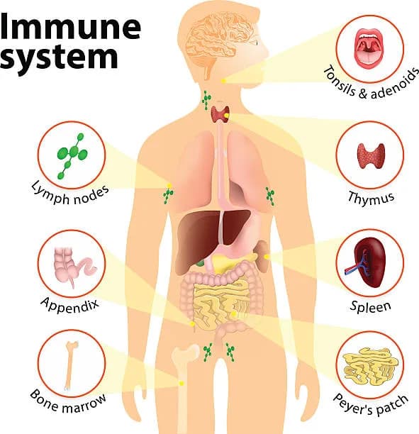
- Bone marrow makes leukocytes (WBC)
- Participate in immune system through hematopoiesis
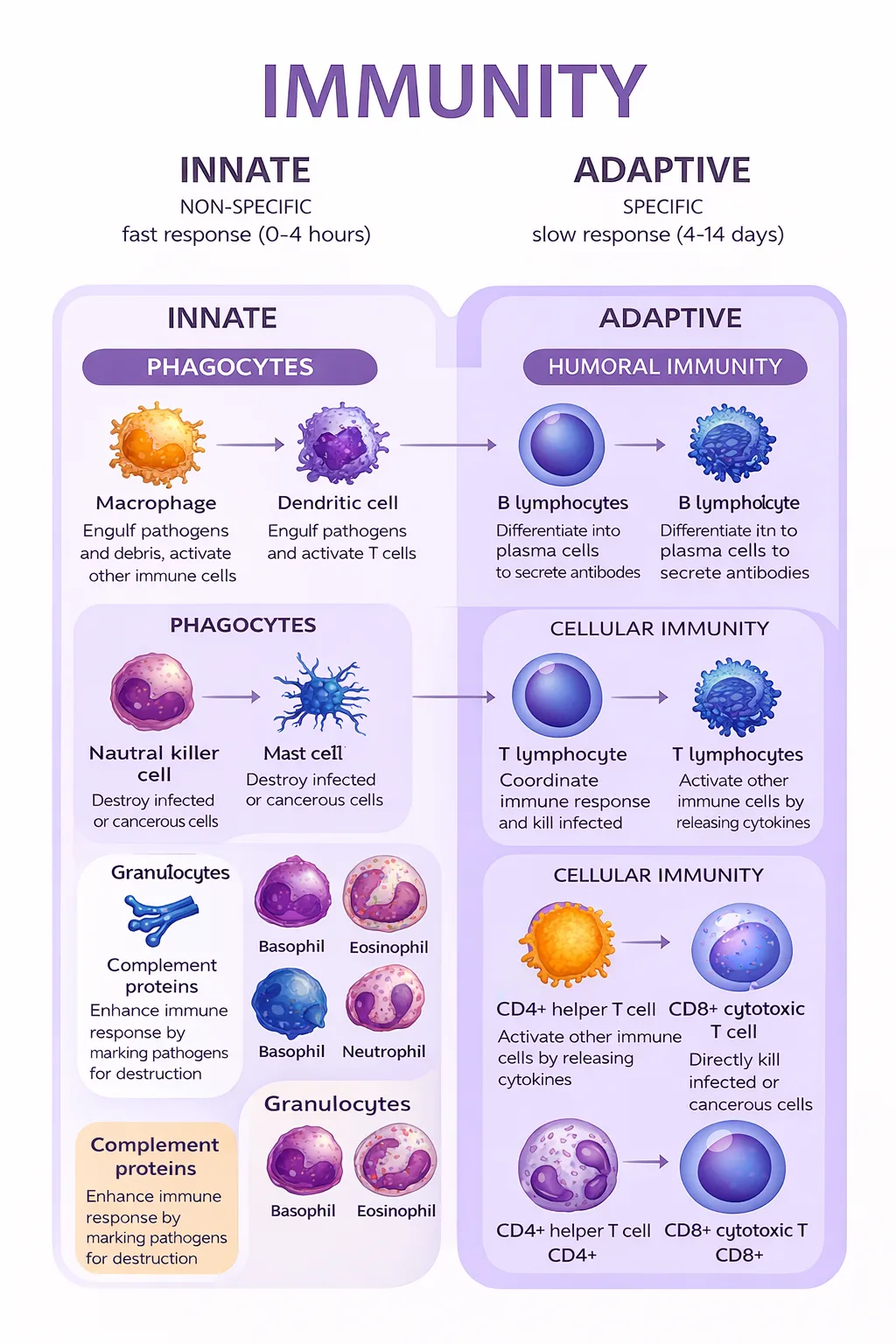
- Granulocytes vs agranulocytes
- Presence/absence of granules in cytoplasm
- Granules contain toxic enzymes/chemicals → released by exocytosis
- Both derive from hematopoietic stem cells
- Granulocytes = neutrophils, eosinophils, basophils
- Agranulocytes = lymphocytes (antibody production, immune modulation, targeted killing of infected cells)
- Lymphocytes
- Monocytes = phagocytic cells in bloodstream (agranulocytes)
- Become macrophages in tissues
- Microglia = CNS
- Langerhans = skin
- Osteoclast = bone
- Become macrophages in tissues
- Spleen = location of blood storage and activation of B cells
- B cells turn into plasma cells to produce antibodies
- When B cells leave bone marrow: mature but naïve (not exposed to antigen yet)
- Humoral immunity (adaptive immunity)
- T cells mature in thymus (small gland in front of pericardium)
- Agents of cell-mediated immunity
- Plasma cells not involved in cell-mediated immunity (not helper T cells)
- Lymph nodes (if swollen, not glands)
- Major component
- Place for immune cells to communicate and mount attack
- B cells activated
- Gut-associated lymphoid tissue (GALT)
- Close to digestive system (site of potential invasion)
- Includes tonsils and adenoids (head), Peyer’s patches (small intestine), lymphoid aggregates in appendix
- Types of immunity
- Nonspecific immune response = without learning
- Specific immune = adaptive learning
- Humoral immunity = driven by B cells and antibodies
- Since B cells from spleen, removal of spleen affects humoral immunity
- Cell-mediated immunity = T cells
8.2 Innate Immune System
Innate immunity includes physical/chemical barriers, complement and interferons, plus immune cells (macrophages, NK cells, granulocytes) that provide rapid nonspecific defense.
- Noncellular nonspecific defenses
- Skin (integument) = first line of defense
- Physical barrier
- Defensins on skin
- Sweat has antimicrobial properties
- Respiratory system
- Cilia + mucus trap stuff
- Uses lysozyme as bacterial enzyme
- GI tract
- Secretes acid to eliminate pathogens
- Gut colonized by bacteria → invaders can’t compete with large population
- Antibiotics reduce gut flora → opportunity for growth of pathogens resistant to antibiotic
- Newborn GI tract uncolonized → susceptible to infection
- Breastmilk has antibodies that help defend
- Skin (integument) = first line of defense
- Complement
- Proteins in blood that act as nonspecific defense
- Classical pathway = binding of antibody to pathogen
- Alternative pathway = no antibodies
- Punch holes in cell walls → unstable
- Interferons
- Infected cells (esp. viral) produce interferons
- Prevent viral replication + dispersion
- Cause nearby cells to decrease production of viral and cellular proteins
- Upregulate MHC class I/II molecules
- ↑ antigen presentation and better detection of infected cells
- Produce flu-like symptoms associated with viral infection
- Cells of innate immune system
- Macrophages
- Agranulocyte
- Derived from monocytes → resident tissue population
- Activated when bacteria enters
- Phagocytize invader via endocytosis
- Digest using enzymes
- Present pieces of invader using MHC
- Carry to cell surface to be recognized by adaptive immune system
- Release cytokines → stimulate inflammation + recruit additional immune cells
- MHC classes
- Class I (CD8)
- On nucleated cells
- Proteins produced in cell loaded + presented on surface
- Allows immune system to monitor cell health
- Endogenous pathway (antigens from inside cell)
- Class II (CD4)
- Displayed by professional antigen-presenting cells
- Exogenous pathway (antigens from outside cell)
- Includes macrophages, dendritic cells (skin), B cells
- Macrophages and dendritic cells have pattern recognition receptors (PRR)
- Toll-like receptors (TLR)
- Recognize category of invader → produce appropriate cytokines to recruit right cell types
- Class I (CD8)
- Natural killer (NK) cells
- Some pathogens downregulate MHC
- NK cells detect downregulation of MHC → induce apoptosis
- Cancer cells downregulate MHC → NK protection
- Granulocytes
- Neutrophils, eosinophils, basophils
- Involved in nonspecific defense
- Neutrophils
- Most populous leukocyte in blood
- Very short-lived
- Phagocytic
- Follow bacteria by chemotaxis (movement via chemical stimuli)
- Detect bacteria once opsonized
- Other cells have receptors for antibodies → attack opsonized cells
- Pus = dead neutrophils
- Neutrophils attack bacteria
- Eosinophils
- Bright red-orange granules
- In allergic reactions and parasites
- Release histamine
- Vasodilation + leakiness of blood
- Allows immune cells to move out of bloodstream into tissue
- Basophils
- Large purple granules
- Allergic responses
- Least populous leukocyte in bloodstream
- Mast cells are related but have smaller granules
- Both release lots of histamine → inflammatory responses
8.3 The Adaptive Immune System
Adaptive immunity uses B cells (humoral antibodies) and T cells (cell-mediated killing/coordination), with clonal selection, memory formation, and MHC-based antigen recognition.
- Cells of the adaptive immune system
- B cells govern humoral response
- T cells govern cell-mediated response
- T cells provide specific defense against pathogens
- Without thymus, could not protect against viruses
- Humoral immunity
- Antibody production
- Takes about a week to become fully effective
- Specific to antigens
- Antibodies can be on cell surface or secreted
- Antibody functions (three possibilities)
- Attract leukocytes to phagocytize antigens immediately (opsonization)
- Agglutinate pathogens into complexes that can be phagocytized
- Neutralize pathogen by blocking invasion of tissues
- Antigen binds antibodies on mast cell surface → degranulation
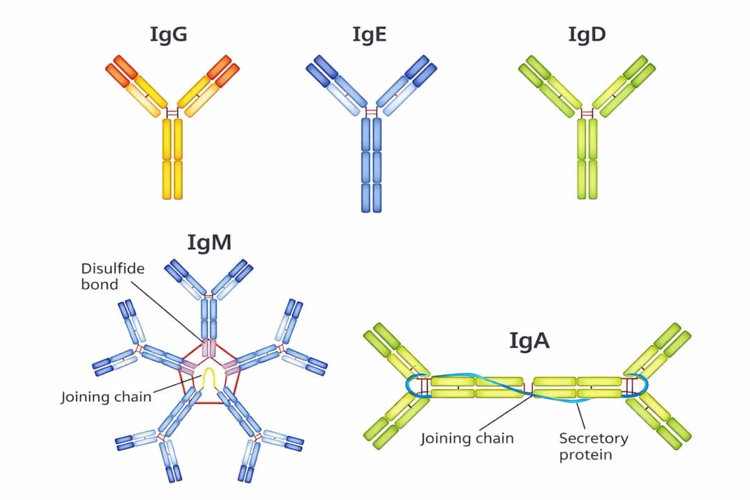
- Antibody structure
- Y-shaped molecules
- 2 identical heavy chains + 2 identical light chains
- Antigen-binding region = variable region (domain) at tips
- Binds only one sequence
- B cells hypermutate antigen-binding region to find best match
- Only high-affinity B cells survive (clonal selection)
- Constant region (domain) = remaining part of molecule
- Many B cells in body → recognize many antigens
- Isotype switching:
- Cytokines stimulate B cells to change isotype of antibody produced
- B cell activation states
- Producing antibodies is energetically expensive
- Naïve B cells wait in lymph node for antigen
- Plasma cells produce large amounts of antibodies
- Memory B cells:
- Wait for re-exposure to same antigen
- Primary response takes 7–10 days
- Memory cells last lifetime of organism
- Secondary response rapid + robust
- Basis of vaccination
- Cytotoxic immunity
- Positive selection:
- Allows maturation of cells that can respond to antigen presented on MHC
- Negative selection:
- Apoptosis in self-reactive cells
- T cell maturation facilitated by thymosin (peptide hormone)
- After leaving thymus: mature but naïve
- Helper T cells (CD4⁺)
- Coordinate immune response
- Secrete lymphokines
- Loss causes HIV; advanced HIV = AIDS
- Weak pathogens can cause devastating consequences
- Respond to antigens on MHC II
- Effective against bacterial, fungal, parasitic
- Cytotoxic T cells (CD8⁺, CTL)
- Kill virally infected cells by injecting toxic chemicals
- Respond to antigens on MHC I
- Most effective against viral
- Suppressor/regulatory T cells
- Express CD4
- Differentiated from helper T cells
- Express Foxp3 protein
- Tone down immune response once infection contained
- Self-tolerance:
- Turns off self-reactive lymphocytes to prevent autoimmune disease
- Memory T cells
- Wait until next exposure
- Robust + rapid response
- Positive selection:
- Activation of the adaptive immune system
- Adaptive and innate systems must work together
- Fight different infections
- Bacterial (extracellular pathogen) infections
- Macrophages are sentinels:
- Engulf bacteria, release inflammatory stuff
- Digest bacteria, create antigens with MHC II
- Cytokines attract neutrophils, macrophages, mast cells
- Immune cells leave bloodstream → travel to infected area
- Dendritic cell travels to lymph:
- Presents antigen to B cells
- B cells with correct antibody proliferate:
- Travel and tag bacteria
- Dendritic cells present antigen to T cells:
- Activate CD4⁺ T cells
- Activates macrophages
- Plasma cells die
- Memory B/T cells remain → faster secondary response
- Macrophages are sentinels:
- Viral infections
- Infected cell makes interferons:
- Reduce permeability of nearby cells
- Reduce viral infection + multiplication
- Causes symptoms
- Infected cells present intracellular proteins via MHC I
- CD8⁺ cells recognize MHC I + antigen complex as foreign:
- Inject toxins → apoptosis
- Infection can be shut down before spread
- NK cells detect absence of MHC I (if downregulated) → apoptosis
- Memory T cells generated
- Infected cell makes interferons:
- Recognition of self and non-self
- Self antigens = normal proteins/carbs on surface of every cell
- Signal cell is not foreign → don’t attack
- If immune system attacks self antigens → autoimmune disorder
- If immune system overreacts to foreign antigen → allergies (hypersensitivity)
- Prevention of autoimmunity:
- T cells educated in thymus, eliminated if respond to self antigen
- Immature B cells that respond eliminated before leaving bone marrow
- If cell survives, glucocorticoids (immunosuppressants) used
- Self antigens = normal proteins/carbs on surface of every cell
- Immunization
- Active immunity:
- Immune system stimulated to produce antibodies against specific pathogen
- Natural exposure = infection triggers B cells to make antibodies
- Artificial exposure = vaccine activates B cells without true infection
- Antigen may be weakened, killed, or part of structure
- Passive immunity:
- Transfer of antibodies to individual
- Only antibodies (not plasma cells) are given
- Ex: placenta transfer, nursing infant
- Post-exposure (rabies, tetanus): immunoglobulins prevent spread
- Active immunity:
8.4 Lymphatic System
The lymphatic system is a one-way vessel network that returns filtered fluid to blood, supports immune surveillance via lymph nodes, and transports dietary fats through lacteals as chyle.
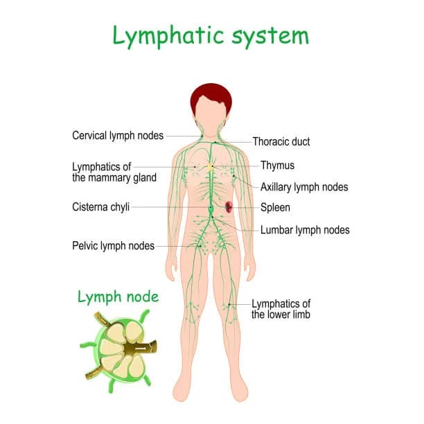
- Structure
- Type of circulatory system
- One-way vessels, larger toward center of body
- Carry lymph
- Join to form thoracic duct (posterior chest)
- Delivers fluid to subclavian vein (near heart)
- Lymph nodes
- Small structures along lymphatic vessels
- Contain lymphatic channel, an artery, and a vein
- Space for immune cells to be exposed to pathogens
- Function
- Equalization of distribution
- Net filtration pressure due to Starling forces
- Lymph vessels drain tissues and return fluid to bloodstream
- If blood has low albumin concentration:
- Less oncotic pressure → more fluid in tissues
- If lymph not blocked → returns fluid
- If lymph overwhelmed → edema
- Transportation of biomolecules
- Lacteals (small lymph vessels) in center of each villus in small intestine
- Chylomicrons (fats) enter lacteals for transport
- Lymph with chylomicrons = milk-white “chyle”
- Immune surveillance
- Lymph nodes are sites where antigen-presenting cells and lymphocytes interact
- B cells proliferate and mature in lymph nodes (germinal centers)
- Equalization of distribution
Spotted something wrong or unclear? Tell us — these notes are revised continuously.