
MCAT Biology · Lesson 3
Embryogenesis and Development
3 min read4 sectionsUpdated
4 sections
3.1 Early Developmental Stages
This section covers the stages from fertilization to early embryonic development, including cleavage, blastulation, gastrulation, and neurulation. It introduces key structures and processes that guide cell differentiation and early tissue formation.
- Fertilization
- Secondary oocyte ovulated on day 14, fertilized 24 hours after ovulation in ampulla
- Sperm penetrates corona radiata and zona pellucida via acrosomal enzymes
- Acrosomal apparatus allows pronucleus entry after meiosis II
- Cortical reaction: Ca²⁺ release depolarizes ovum, preventing polyspermy
- Fertilization membrane: depolarized, impenetrable
- Twins
- Dizygotic: 2 eggs + 2 sperm, genetically like siblings
- Monozygotic: single zygote splits, may result in conjoined twins
- Classified by shared structures (amnion, chorion)
- Cleavage
- Rapid mitotic divisions as zygote travels to uterus
- ↑ N:C ratio and surface area to volume ratio
- Indeterminate cleavage: cells can become complete organism
- Determinate cleavage: cells committed to specific fate
- Blastulation
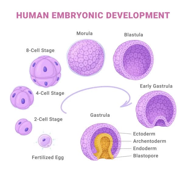
- Zygote → morula → blastocyst → gastrula
- Blastocyst: trophoblast → chorion/placenta; inner cell mass → organism
- Blastocoel: fluid-filled cavity
- Implantation: blastula burrows into endometrium
- Trophoblast forms chorionic villi for maternal-fetal gas exchange
- Umbilical cord connects embryo to placenta
- Yolk sac supports embryo before placenta functions
- Allantois: early fluid exchange, contributes to umbilical cord
- Amnion: thin membrane, amniotic fluid as shock absorber
- Gastrulation
- Formation of primary germ layers via membrane invagination (archenteron)
- Blastopore:
- Deuterostomes → anus
- Protostomes → mouth
- Germ layers:
- Ectoderm: epidermis, hair, nails, epithelia, nervous system, inner ear, eye, adrenal medulla
- Mesoderm: muscles, circulatory, excretory, gonads, connective tissue, adrenal cortex
- Endoderm: epithelial linings of digestive/respiratory tracts, lungs
- Differentiation
- Selective transcription → cell-specific gene expression
- Induction: cells influence fate of nearby cells
- Guides axon development
- Neurulation
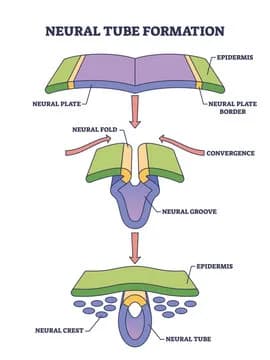
- Mesodermal notochord induces ectoderm to form neural folds → neural tube (CNS)
- Neural crest cells → PNS and other tissues
- Ectodermal cells migrate over neural tube
- Failure → spina bifida, anencephaly
- Problems in early development
- Teratogens: alcohol, drugs, viruses, chemicals
- Maternal physiology influences development (e.g., hyperglycemia)
3.2 Mechanisms of Development
Discusses cell specialization, differentiation, and communication. Introduces stem cell potency, apoptosis, cell migration, and regenerative capacity.
- Cell specialization
- Cells perform distinct functions (e.g., pancreas: enzymes vs hormones)
- Specification/Determination
- Specification: reversible
- Determination: irreversible commitment
- Morphogens guide determination
- Differentiation
- Structural, functional, biochemical changes
- Stem cells: embryonic and adult tissues
- Potency:
- Totipotent → any cell type
- Pluripotent → any except placental
- Multipotent → group-specific
- Cell-to-cell communication
- Induced/responsive cells must be competent
- Signal types:
- Autocrine → same cell
- Paracrine → nearby cell
- Juxtacrine → adjacent cell via receptors
- Endocrine → hormones
- Inducers
- Growth factors, act on specific cells
- Reciprocal development
- Morphogen gradients influence fate
- Cell migration
- Cells detach and move (e.g., neural crest cells)
- Cell death
- Apoptosis: programmed, forms blebs → phagocytes recycle
- Necrosis: injury-induced, substances released
- Regeneration
- Complete: identical replacement
- Incomplete: non-identical replacement
- Human regeneration varies: liver high, heart low, kidney moderate
- Senescence and aging
- Cells stop dividing after ~50 cycles
- Shortened telomeres, telomerase can maintain divisions (cancer survival)
3.3 Fetal Circulation
Explains fetal circulation adaptations, placental function, and maternal-fetal blood separation.
- Maternal/fetal blood separate, HbF has ↑ O₂ affinity
- Placenta gas exchange
- Umbilical cord: 2 arteries (to placenta), 1 vein (from placenta)
- Placenta produces P, E, hCG; antibodies cross
- Fetal liver/lungs inactive, detox via maternal liver
- 3 shunts bypass lungs and liver:
- Lung: foramen ovale (RA → LA), ductus arteriosus (pulm artery → aorta)
- Liver: ductus venosus (placental blood → IVC)
3.4 Gestation and Birth
Describes gestation timeline, fetal development by trimester, and parturition process.
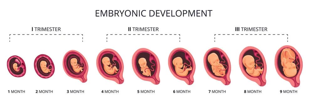
- Gestation: 280 days, 3 trimesters, longer in larger animals
- First trimester
- Major organs form; heart beats at 22 days
- By 8 weeks: most organs formed, brain developed, embryo → fetus (~9 cm)
- Second trimester
- Rapid growth, human facial features
- Length: 30–36 cm
- Third trimester
- Further growth and brain development
- Antibody transfer, reduced activity
- Birth
- Vaginal childbirth: uterine contractions (oxytocin, prostaglandins)
- Cervix thins, amniotic sac ruptures
- Delivery of fetus
- Placenta and umbilical cord expelled (afterbirth)
Spotted something wrong or unclear? Tell us — these notes are revised continuously.