
MCAT Biology · Lesson 2
Reproduction
4 min read3 sectionsUpdated
Jump to a section3
3 sections
2.1 Cell Cycle and Mitosis
This section covers the basics of the cell cycle, including interphase, mitosis, and checkpoints. Key concepts include regulation by cyclins and CDKs, the role of p53 in DNA damage response, and the phases of mitosis that ensure identical daughter cells.
Prototherians and Metatherians
- Prototherians (platypus, echidna): lay hard-shelled amniotic eggs (oviparity).
- Metatherians (marsupials like koalas, kangaroos): fetus develops in uterus, then moves to marsupium (pouch).
Cell Ploidy
- Animal autosomal cells: diploid (2n)
- Germ cells: haploid (n)
Cell Cycle
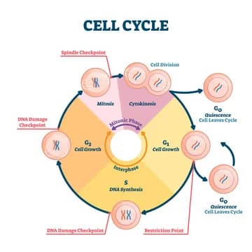
- Interphase: G1, S, G2 (90% of cycle)
- G0: non-dividing, no preparation for division
- G1 (Pre-synthetic gap): organelle production, cell growth; checkpoint prevents progression if DNA damaged.
- S (Synthesis stage): DNA replication; chromosome has 2 identical chromatids (ploidy unchanged: still 46 chromosomes, 92 chromatids).
- G2 (Post-synthetic gap): checkpoint ensures organelles, cytoplasm, and DNA replication readiness.
- M Stage (Mitosis): division of nucleus and cytoplasm (cytokinesis).
Control of Cell Cycle
- G1/S checkpoint ensures DNA integrity.
- p53 protein: key regulator.
- Cyclins and CDKs: cyclin-CDK complexes phosphorylate TFs to drive cell cycle progression.
Cancer
- Mutation in TP53 → unchecked division.
- Tumors: rapid division; metastasis: spread to other tissues.
Mitosis Phases
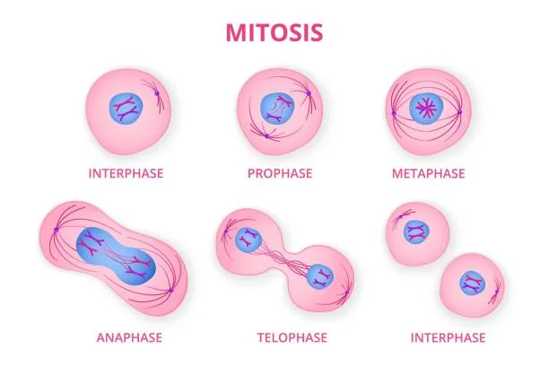
- Prophase: chromatin condenses; centrioles separate → spindle fibers; nuclear membrane dissolves; kinetochores form.
- Metaphase: chromosomes align at metaphase plate; spindle fibers attach.
- Anaphase: centromeres split; sister chromatids move to poles.
- Telophase & Cytokinesis: spindle disappears; nuclear membrane reforms; chromosomes uncoil; cytoplasm divides.
- Finite divisions: 20–50 in humans.
2.2 Meiosis
Meiosis produces four nonidentical haploid gametes through one round of DNA replication and two rounds of division. Homologous chromosome separation in Meiosis I and sister chromatid separation in Meiosis II generate genetic diversity through recombination and independent assortment.
General
- Occurs in gametocytes (germ cells)
- 1 replication, 2 divisions
- Meiosis I: reductional division (homologous chromosomes separate)
- Meiosis II: equational division (similar to mitosis)
Meiosis I
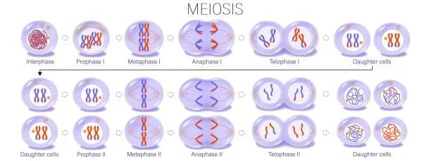
- Prophase I: chromatin condenses; spindle forms; homologous chromosomes synapse → tetrads; crossing over occurs at chiasmata (between homologs, not sisters) → genetic recombination.
- Metaphase I: tetrads align at metaphase plate; homologs opposite each other.
- Anaphase I: homologous pairs separate (disjunction); segregation ensures paternal/maternal chromosomes separate.
- Telophase I: nuclear membrane reforms; cytokinesis → haploid cells.
- Interkinesis: short rest; chromosomes may partially uncoil.
Meiosis II
- Analogous to mitosis; sister chromatids separate.
- Results in 4 haploid gametes.
2.3 Reproductive System
This section outlines male and female reproductive anatomy, gametogenesis, sexual development, and the menstrual cycle. Emphasis is on gamete formation, hormone regulation, and physiological changes across life stages including puberty, pregnancy, and menopause.
Sex Determination
- 23rd chromosome pair: XX female, XY male
- Males: hemizygous X → express X-linked traits
- Y chromosome: SRY gene → testis formation
- Absence of Y → female
Male Reproductive System (SEVE(N) UP)
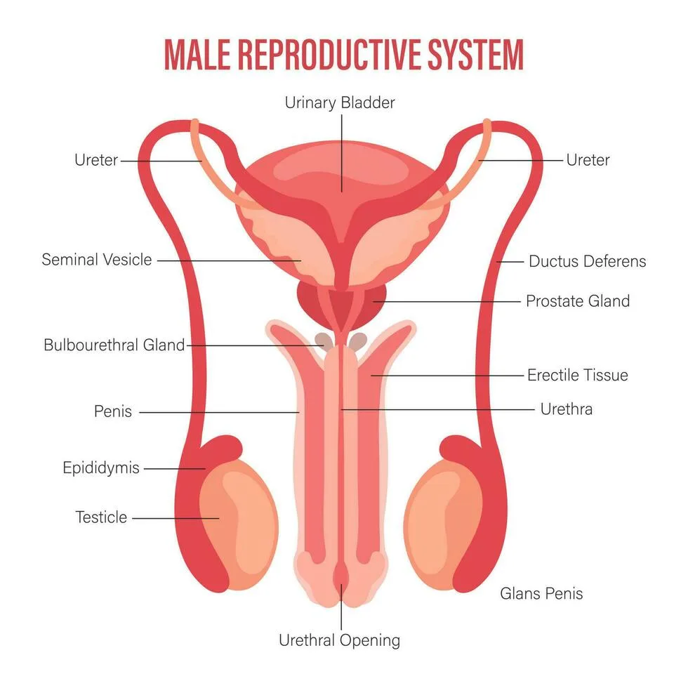
- Seminiferous tubules → Epididymis → Vas deferens → Ejaculatory duct → Nothing → Urethra → Penis
Male Anatomy & Spermatogenesis
- Testes: seminiferous tubules (sperm), interstitial cells of Leydig (T production)
- Sperm maturation in epididymis
- Ejaculation: vas deferens → ejaculatory duct → urethra; mixes with seminal fluid (seminal vesicles, prostate, bulbourethral gland)
- Spermatogenesis: spermatogonia → primary spermatocytes → secondary spermatocytes → spermatids → spermatozoa
- Structure: head (DNA, acrosome), midpiece (ATP), flagellum (motility)
- ~3 million sperm/day
Female Anatomy & Oogenesis
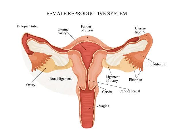
- Ovaries: produce E & P; contain follicles
- Ovulation: one egg/month → fallopian tube → uterus
- Oogenesis: all oogonia formed fetal → primary oocytes at birth → meiosis I at menarche → secondary oocyte + polar body
- Secondary oocyte arrested in metaphase II until fertilization
- Layers: zona pellucida (glycoproteins), corona radiata (outer cell layer)
Sexual Development
- Hypothalamus releases GnRH → anterior pituitary releases FSH/LH → triggers sex hormones
- Male: fetal androgen, puberty ↑T, FSH → Sertoli cells, LH → Leydig cells
- Female: FSH → E, E → reproductive system & secondary sexual characteristics, P → endometrium maintenance
- Negative feedback loops regulate hormone levels
Menstrual Cycle
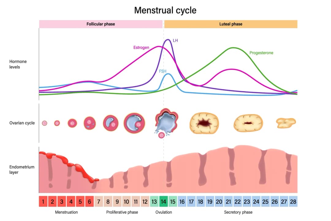
- Follicular Phase: menstruation, low E/P → ↑GnRH → ↑FSH/LH → follicle development → E ↑ → endometrium regrows
- Ovulation: E peak → positive feedback → GnRH/FSH/LH surge → ovulation
- Luteal Phase: corpus luteum forms → P ↑ → negative feedback → prevent multiple ovulations; supports endometrium
- Menstruation: if no implantation → corpus luteum degenerates → P ↓ → endometrium sloughs off → cycle restarts
Pregnancy
- Fertilization → zygote → blastocyst → implantation
- hCG maintains corpus luteum early; placenta takes over hormone production later
Menopause
- Ovarian sensitivity ↓ → endometrium atrophies → menstruation stops
- FSH/LH ↑ due to loss of negative feedback
- Occurs ~45–55; physical and mental changes
Spotted something wrong or unclear? Tell us — these notes are revised continuously.