
MCAT Biology · Lesson 11
Musculoskeletal System
4 min read2 sectionsUpdated
Jump to a section2
2 sections
11.1 Muscular System
This section covers the 3 muscle types and how skeletal muscle structure (sarcomeres, bands, SR/T-tubules) supports contraction, plus key ideas in twitch, summation/tetanus, and fatigue/O₂ debt.
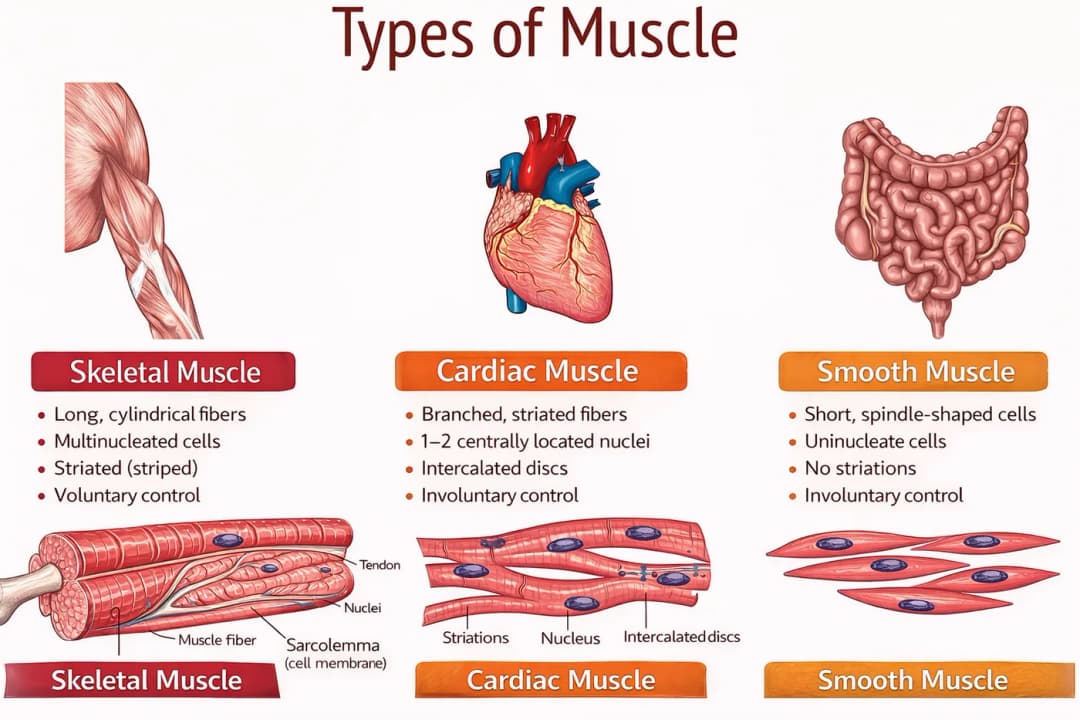
- Types of muscle
- Skeletal
- Voluntary movement, somatic nervous system
- Sarcomeres → striated
- Red fiber = slow twitch
- High myoglobin, high mitochondria, aerobic
- White fibers = fast twitch
- Low myoglobin (less iron so lighter)
- Sustained = red, rapidly = white
- Smooth
- Involuntary, more sustained contractions
- Maintains constant tension (blood vessels), tonus
- Can contract with myogenic activity
- Cardiac muscle
- Characteristics of both
- Connected with intercalated discs, containing gap junctions
- Myogenic activity = maintain over rhythm
- Starts at SA node → AV node → bundle of His → Purkinje fibers
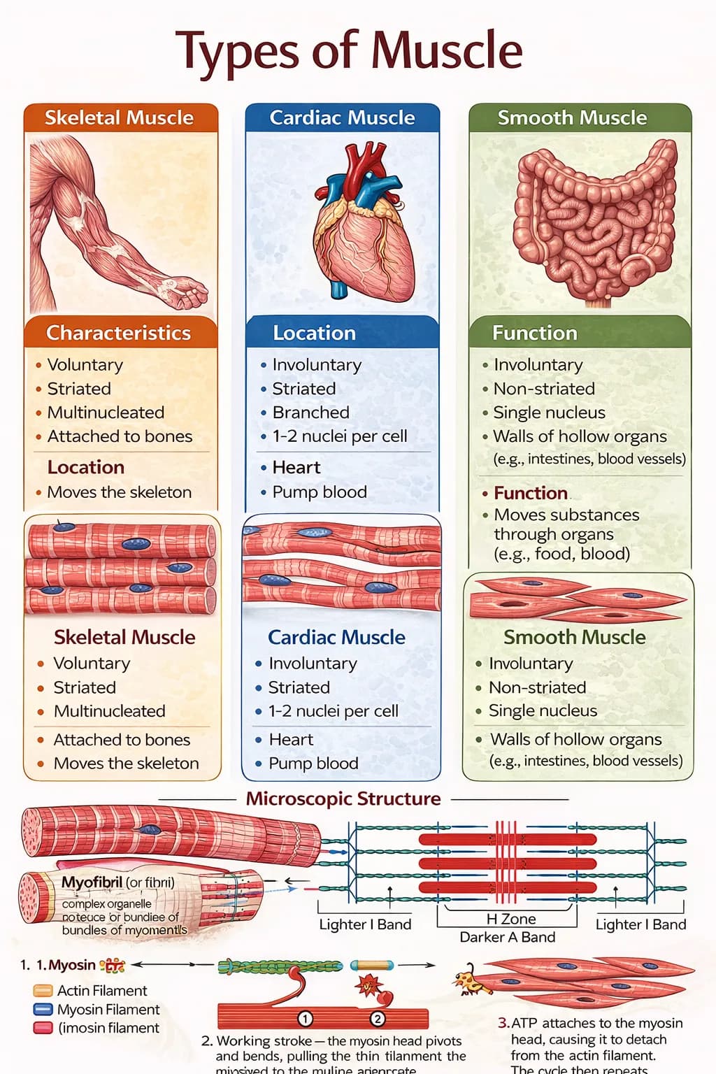
- Skeletal
- Microscopic structure of skeletal muscle
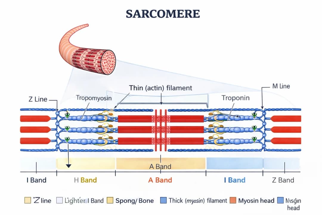
- Sarcomere
- Thick = myosin
- Thin = actin (troponin, tropomyosin)
- Titin acts as a spring and anchors actin and myosin filaments together
- Z line defines boundaries of sarcomere
- M line runs down center
- I band only has thin filaments
- H zone has thick
- A band thick
- Z end of alphabet = end of sarcomere
- Mnemonic
- M = middle of the myosin
- I = thin letter
- H = thick letter
- A = all of thick filament
- Sarcomere
- Gross structure of myocytes
- Myofibrils surrounded by covering from SR, has high [Ca]
- Cell membrane is sarcolemma
- Propagates AP and distributes
- Uses T tubules, perpendicular to myofibrils
- Muscle fiber is multiple myofibrils = myocyte
- Muscle contraction
- Initiation
- Contraction at NMJ, signal travels to nerve terminal, motor end plate
- Nerve terminal is motor end plate
- Nerve terminal and myocyte is motor unit
- Ca released from SR, binds to troponin → changes tropomyosin, troponin
- Shortening of the sarcomere
- Myosin hydrolyzed ATP, binds with site
- ATP releases myosin from actin, sliding filament mechanism
- Relaxation
- ACh degraded by acetylcholinesterase, termination of signal
- ATP binds to myosin and releases
- Sarcomere returns to original width
- Initiation
- Stimulation, summation, muscle fatigue
- Simple twitch
- Single muscle fiber to brief stimulus at or above threshold
- Latent period is time between reaching threshold and onset of contraction
- Muscle contracts, assume Ca cleared from sarcoplasm
- Summation and tetanus
- Insufficient time to relax, if frequent and prolonged stimulation
- Frequency summation
- If too much, then tetanus
- Oxygen debt and muscle fatigue
- Require ATP, need oxygen to generate
- Creatine phosphate transferring phosphate group ATP
- Creation reversed during muscle use
- Red muscles switch to anaerobic metabolism to make lactic acid
- Difference in O₂ needed and present is O₂ debt
- Metabolize lactic acid after exercise
- Most goes to pyruvate
- Simple twitch
11.2 Skeletal System
This section outlines axial vs appendicular skeleton, bone/cartilage composition and structure (macro + micro), remodeling hormones, ossification types, and how joints/muscles produce movement.
- Skeletal structure
- Axial is skeleton, skull, ventral column, rib bone, hyoid bone
- Appendicular is upper limb
- Bone composition
- Connective tissue, from embryonic mesoderm
- Bone is harder than cartilage
- Macroscopic bone structure
- Strength comes from compact bone
- Spongy (cancellous) bone
- Lattice has trabeculae
- Cavities between are bone marrow
- Red is hematopoietic stem cells, make cells in blood
- Yellow marrow is fat, inactive
- Appendicular skeleton has long bones
- Shafts are diaphysis
- Swell at end to metaphysis
- Terminate in epiphysis
- Epiphysis have spongy bone to disperse force and pressure
- Internal edge is epiphyseal (growth) plate
- Mitotic cells for growth in puberty, close at adulthood
- Periosteum around long bone to protect and site for muscle attachment
- Tendons = muscle to bone
- Ligaments = bones together at joints
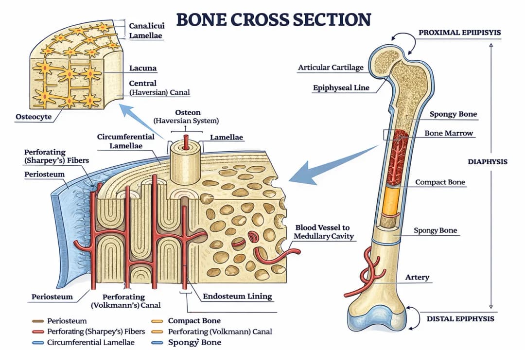
- Microscopic bone structure
- Strength from bone matrix
- Organic components = collagen, glycoproteins, peptides
- Inorganic = Ca, PO₄, OH → make hydroxyapatite crystals
- Strong bones have distribution of materials
- Units called osteons
- Concentric circle of bony matrix called lamellae surrounding central channel
- Longitudinal = haversian canals
- Transverse = Volkmann's canals
- Lacunae between lamellar rings
- Have bone cells known as osteocytes
- Canaliculi interconnective to tiny channels
- Allow for exchange of nutrients
- Strength from bone matrix
- Bone remodeling
- Osteoblasts build bone, osteoclasts resorb it
- Secrete bone matrix
- Turnover of bone, resorption releases stuff back into blood
- Remodels in relation to stress
- Parathyroid hormone released by parathyroid glands in response to low blood Ca
- Vitamin D activated, promotes resorption
- Calcitonin promotes bone formation
- Cartilage
- Firm but elastic matrix called chondrin, secreted by chondrocytes
- Fetal skeleton mostly cartilage because they are elastic
- Ossification
- Endochondral = formation of long bones
- Intramembranous = connective tissue transformed into, replaced by bone (skull)
- Joints and movement
- Immovable joints = sutures
- Movable joints = hinge
- Strengthened by ligaments
- Synovial capsule, enclosed joint capsule
- Synovium secretes synovial fluid, lubricates
- Articular cartilage coats surface so impact is restricted to cartilage and not bone
- Lack of cartilage = arthritis = bones rubbing on each other
- Contraction
- End of muscle larger attachment to bone is origin (proximal)
- Smaller attach is insertion (distal)
- Muscles are antagonistic
- Can also be synergistic
- Muscle kinds
- Flexor = decreases angle (biceps)
- Extensor = increases angle (triceps)
- Abductor moves part of body away from midline (deltoid)
- Adductor moves toward midline (pectorals)
- Medial and lateral rotation (motions in limbs)
Spotted something wrong or unclear? Tell us — these notes are revised continuously.